Introduction
Isolated epispadias represents the mildest anomaly within the exstrophy–epispadias complex. It is an uncommon condition, with an estimated incidence of 1 in 120,000 male births [1]. In more than 90% of cases, epispadias occurs in association with bladder exstrophy [2,3]. The incidence of epispadias with an intact or phimotic prepuce remains unknown, as reports are limited to isolated cases. To date, 22 cases of epispadias with an intact prepuce have been documented—14 cases summarized in a 2016 review [4] and 8 additional cases reported in a 2014 single-center series [5]. Furthermore, 8 cases of epispadias with a phimotic intact prepuce have been described in the literature [6-13]. The present case represents the 23rd reported instance of epispadias with an intact prepuce and the 9th involving a phimotic intact prepuce. Although it may not be a rare condition, it is often underreported because it can be easily missed.
This report details a case of penopubic epispadias with an intact prepuce in a 6-month-old male infant, initially misdiagnosed as phimosis post-circumcision. The authors also reviewed existing literature concerning the condition’s presentation and surgical treatment.
Case presentation
A 6-month-old male infant, who had a traditional circumcision at one month of age, was brought to our hospital due to phimosis. He had no reported history of urinary tract infections or abnormal urinary flow. His growth and development were on track for his age, and a review of his overall health showed no significant findings. The pregnancy and delivery were supervised and uneventful; the child was full-term.
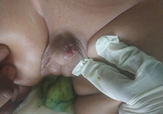
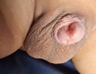
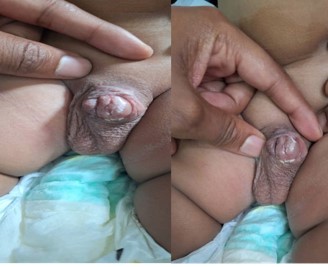
During a surgical exploration to correct phimosis, the infant was unexpectedly diagnosed with penopubic epispadias and an intact prepuce. The prepuce was severely phimotic, preventing retraction and visualization of the glans.
After retracting the prepuce under anesthesia, the condition of penopubic epispadias with a broad, healthy urethral plate was revealed. There were no signs of meatal stenosis. The penis had mild chordea and appeared short and was entirely covered by the intact prepuce. Both testes were normally sized and consistently felt within the scrotum. The diagnosis of penopubic epispadias was confirmed, and the planned circumcision was postponed.
Laboratory investigations, including urinalysis, renal function tests, and abdominal ultrasound, were within normal limits.
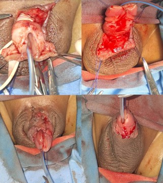
After thorough parental counseling, the infant underwent a Modified Cantwell-Ransley repair for epispadias one-month post-presentation. The surgical procedure involved successful urethral tubularization, corporal rotation, and neomeatus creation, with an uneventful operative and postoperative course. The urethral catheter was removed on postoperative day seven.
At the 3-month follow-up, the repair remained stable, with an aesthetically acceptable penile appearance and effective voiding through the neomeatus in a single stream, consistent with previously reported outcomes [1,14,5].
Discussion
Epispadias, a malformation that constitutes less than 10% of the exstrophy-epispadias spectrum, typically presents with an obvious dorsal meatus and underdeveloped prepuce at birth [16]. However, when the prepuce appears normal, as in this patient, the condition can be concealed, leading to a delayed diagnosis [2,14].
The urethra and prepuce develop in tandem; a urethral developmental defect often leads to a corresponding failure in prepuce development. The pathogenesis of the exstrophyepispadias complex is attributed to defective cloacal membrane development. Urethral embryogenesis begins in the second intrauterine month with the cloaca’s division into the posterior anorectal canal and the anterior primitive urogenital sinus. The urogenital sinus develops into the bladder, proximal prostatic urethra, and membranous urethra. Distal to this, the phallic cloaca extends through the genital tubercle, which, through proliferation, moves caudally to the developing glans. Failure in this latter step results in epispadias [1,14].
The development of the prepuce commences in the third month of gestation with the proliferation of the genital tubercle. Subsequently, following the median cleavage of the urethral plate, two tissue fold sets emerge on the ventral surface, flanking the urethral groove. The medial endodermal folds coalesce in the ventral midline, forming the urethra, while the lateral ectodermal folds merge over the developing urethra to create the penile shaft skin and the prepuce. An ectodermal ring forms just proximal to the glans penis, advancing over its corona to fully enclose it as the prepuce or foreskin. Another perspective suggests that the prepuce arises from a blend of preputial folding and the inward growth of a cellular lamella, which constructs the prepuce, glans, corona, and coronal sulcus mucosa [1,14].
The presence of epispadias with an intact prepuce is attributed by McCahill et al. to the theory of active mesenchyme growth between the preputial fold and the glandular lamella, which pushes the fold distally to fully cover the glans. In instances where these folds originate proximal to the urethral defect, they envelop both the deficient urethra and the glans, thereby precluding the epispadias from affecting prepuce development. Nonetheless, this theory does not adequately explain cases of proximal epispadias, and despite the formulation of various alternative hypotheses, the simultaneous development of epispadias and an intact prepuce remains difficult to account for [1,6,14].
Epispadias, when isolated, can manifest as glanular, coronal, shaft, or penopubic. While distal forms rarely involve incontinence, penopubic epispadias features a urethral meatus extending to the membranous urethra, which can lead to bladder neck and sphincter insufficiency [1].
Epispadias typically involves incomplete tubularization of the urethral plate, leading to a dorsal urethral meatus. Although a deficient or abnormal dorsal prepuce often accompanies epispadias, simplifying diagnosis, the presence of an intact prepuce is exceptionally rare. Consequently, these cases might go undetected until later or be found incidentally during circumcision or phimosis assessment. Clinical indicators for this variant include a broad, spade-like glans, a dorsally oriented preputial opening, a potentially palpable gap between the corpora cavernosa, dorsal chordee, and irregularities in the penile raphae [1].
An unusual presentation of epispadias can be phimosis. In some reported instances, the condition was only identified during intraoperative circumcision or surgical exploration [1,613,14,16].
Surgical technique
The surgical objective is to reconstruct the epispadiac urethra and glans, while also correcting any dorsal chordee. The specific surgical technique employed for reconstruction is dictated by the severity of the epispadias [1,2,14,16].
For coronal, shaft, or penopubic epispadias, the urethra is repositioned between the corpora cavernosa utilizing the Cantwell-Ransley technique. The Modified Cantwell-Ransley technique continues to be a dependable method for primary penopubic epispadias repair, yielding positive cosmetic and functional results, as evidenced in this case and documented in other studies [1,14,15]. A U-shaped incision was created, with its limbs positioned at the border of the urethral plate and the curve at the epispadias’ proximal end. The urethral plate was then tubularized around a 10F urethral catheter using Vicryl 6/0 suture. Following this, the two corporal bodies were brought together over the reconstructed urethra, and the prepuce was repaired.
For glandular epispadias, the IPGAM procedure effectively yields favorable results [1,14].
Alternatively, the Complete Penile disassembly technique, as proposed by Mitchell, is an option when a hypospadias urethra needs to be created. The success of this method is evaluated by the cosmetic outcome of the penile reconstruction, the preservation of erectile function, and the achievement of urinary continence [1].
Penile lengthening is carried out if the corpora are not adequately fixed to the anterior aspect of the pubic bones. This procedure involves extending the penis and securing the corpora cavernosa to the anterior side of the pubic bones, flanking the symphysis, using non-absorbable sutures. Excess pre-symphysis fat is excised, and the dorsal skin is elongated for aesthetic enhancement [1,14].
Boys diagnosed with epispadias and an intact prepuce tend to experience fewer complications, require less follow-up intervention, and demonstrate superior rates of urinary continence [14].
This case emphasizes the need for a high index of suspicion when circumcision is requested for children with unusual penile anatomy. Circumcision performed without a comprehensive genital examination could potentially hinder subsequent reconstructive procedures [1,6-13,14,16].
Conclusion
Epispadias with an intact prepuce presenting as phimosis is extremely rare and may result in delayed diagnosis. Thorough evaluation of the genital anatomy before circumcision is essential to prevent misdiagnosis and unintended surgical intervention.
Declarations
Statements
1. Written informed consent was obtained from the patient’s parents for publication and any accompanying images.
2. All authors attest that they meet the current ICMJE criteria for authorship.
Funding: We declare there was no external source of funding.
Competing interest: All authors declare no competing interests.
Abbreviations: ICMJE: International Committee of Medical Journal Editors; IPGAM: Inverted Preputial Glanuloplasty and Meatoplasty.
References
- Sarma VP. Concealed epispadias: a rare anomaly—case report and review. Int Surg J. 2019; 6: 2979–81.
- Waziri AM, Abubakar BM, Adamu S, et al. Epispadias in a child with intact prepuce: a rare congenital abnormality. Open J Urol. 2016; 6: 19–22.
- Maitama HY, Ahmed M, Bello A, Mbibu HN. Epispadias with complete prepuce: a rare anomaly. Afr J Urol. 2012.
- Garge S. Isolated epispadias with intact prepuce: a review. Afr J Paediatr Surg. 2016; 13: 84–6.
- Arshad AR, et al. Epispadias with intact prepuce: a single-center experience. Afr J Paediatr Surg. 2014; 11: 322–5.
- McCahill PD, Leonard MP, Jeffs RD. Epispadias with phimosis: an unusual variant of the concealed penis. Urology. 1995; 45: 164–8.
- Merlob P, Mor N, Reisner SH. Epispadias with complete prepuce and phimosis in a neonate. Clin Pediatr (Phila). 1987; 26: 43–5.
- Gearhart JP, Jeffs RD. Exstrophy-epispadias complex and bladder anomalies. In: Wein AJ, Kavoussi LR, Partin AW, Peters CA, editors. Campbell-Walsh Urology. 10th ed. Philadelphia: Saunders; 2012. p. 3315–48.
- Shapiro E, Lepor H, Jeffs RD. The inheritance of the exstrophyepispadias complex. J Urol. 1984; 132: 308–10.
- Ebert AK, Reutter H, Ludwig M, Rösch WH. The exstrophy–epispadias complex. Orphanet J Rare Dis. 2009; 4: 23.
- Raghavaiah NV. Isolated epispadias with phimosis. Br J Urol. 1976; 48: 303.
- Krishna A, Iyer KR. Isolated epispadias with intact prepuce: a case report. Indian Pediatr. 1987; 24: 599–600.
- Bos EME, van der Toorn F, de Jong TPVM. Isolated epispadias with intact prepuce: a report of three cases. J Pediatr Urol. 2014; 10: 1132–5.
- Garge S. Concealed epispadias: report of two cases and review of literature. Urology. 2016; 90: 164–8.
- Kaya M, Sancar S, Ozcakir E. Epispadias with intact prepuce: a case report. Pediatr Urol Case Rep. 2015; 2: 23–8.
- Bos EM, Kuijper CF, Chrzan RJ, Dik P, Klijn AJ, de Jong TP. Epispadias in boys with an intact prepuce. J Pediatr Urol. 2014; 10: 67–73.