Introduction
Initially developed for the management of hypertension in
the 1970s, Minoxidil is now widely used to treat hair loss in both
men and women. Functioning as an active vasodilator, Minoxidil promotes blood flow by relaxing blood vessels [1]. It is currently accessible Over The Counter (OTC) as topical solutions of
2% and 5% and is prescribed as a daily oral medication for the
management of hypertension or androgenic alopecia [2].
The most notable side effects of the topical formulation of
Minoxidil are scalp irritation or allergic contact dermatitis. Systemic side effects include tachycardia, increased cardiac function and stroke volume, sodium retention, and abnormal hair
growth [3]. Information leaflets and drug monographs list the
main ocular side effect as conjunctivitis and blurred vision,
mostly associated with the topical formulation of Minoxidil.
Nonetheless, there have been a few case reports that have previously shown Minoxidil-induced ocular pathologies, all associated with the use of its topical formulation [3-6]. Herein, we
present a case of Central Retinal Vein Occlusion (CRVO) with
secondary Cilioretinal Artery Occlusion (CLRAO), likely caused
by the chronic use of Minoxidil.
Case presentation
A 35-year-old male presented to the Royal Victoria Hospital
(RVH) Emergency Department (ED) with sudden vision loss in
the superior nasal quadrant of the right eye (OD). He reported no significant medical history other than alopecia and migraines. His family history was non-contributory, and his only
prescription medications were nortriptyline 10 Milligrams (mg)
daily and Minoxidil 1.25 mg daily.
The initial ophthalmic evaluation showed his Best-Corrected
Visual Acuity (BCVA) to be 20/40 OD and 20/20 in the left eye
(OS). The maximum intraocular pressure (Tmax) was 15 OD
and 19 OS. His pupils were equal, symmetric, and reactive to
light, with no Relevant Afferent Pupillary Defect (RAPD), and he
had full Extraocular Motion (EOM) in both eyes (OU). Slit-lamp
biomicroscopy examination of the anterior segment was unremarkable. However, Dilated Fundus Examination (DFE) of the
posterior segment revealed scattered mild retinal hemorrhages
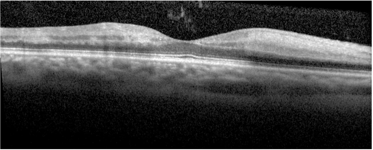
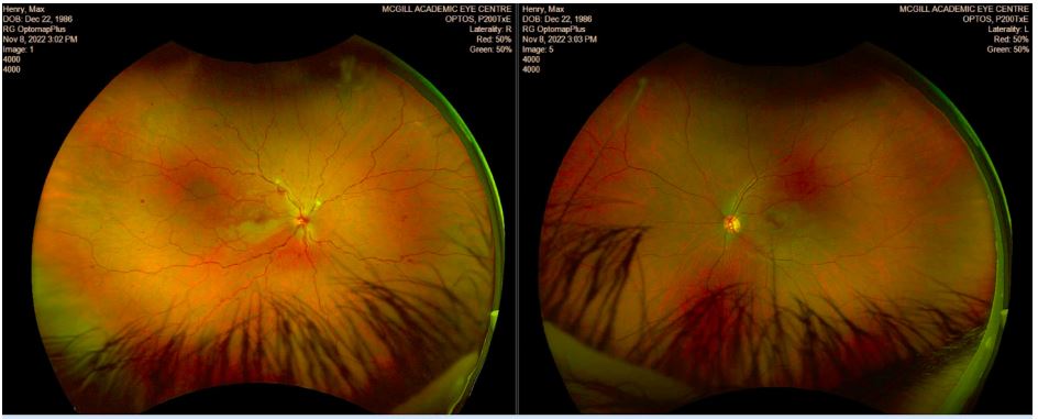
and tortuosity with patchy retinal whitening in the inferior macula (Figure 1A). Optical Coherence Tomography (OCT) revealed
inner retinal hyper reflectivity (Figure 2). He was clinically diagnosed with CRVO with secondary CLRAO.
Fluorescein Angiography (FA) performed two days later revealed a small peripheral branch with delayed filling and mild
vascular leakage, in keeping with CRVO. However, the CLRA filling was found to be within normal limits. Fundus photography
at day six revealed macular edema, dot-blot hemorrhages, and
venous tortuosity (Figure 3). At this time, he was advised to
cease the use of Minoxidil. He was also treated with a course
of prednisone, tapered from 50 mg orally for 5 days between
weeks two and three. Over the next few weeks to months, the
patient underwent detailed systemic evaluations and investigations. An Electrocardiogram (EKG) and Transthoracic Echocardiogram (TTE) were negative for cardioembolic events. Computed Tomography (CT) of the head without Contrast (C-) showed no evidence of acute territorial infarct, intracranial hemorrhage,
mass lesion, or hydrocephalus, and CT Angiography (CTA) of the
neck revealed no acute occlusion or significant stenosis along
the cervical and intracranial arterial vasculature. Importantly,
bilateral ophthalmic arteries were patent. Additional investigation with Magnetic Resonance Imaging (MRI) with contrast
of the head and orbits was negative for any ischemic lesion or
optic neuropathy. Investigations for infectious causes, such as
Bartonella henselae, toxoplasmosis, and Mycobacterium tuberculosis, were also negative. Investigations for inflammatory
causes, such as antinuclear antibodies, protein C, antithrombin,
and anti-neutrophil cytoplasmic antibodies, were all negative.
Furthermore, genetic analysis for factor V and prothrombin
(20210 A) gene mutations were also negative.
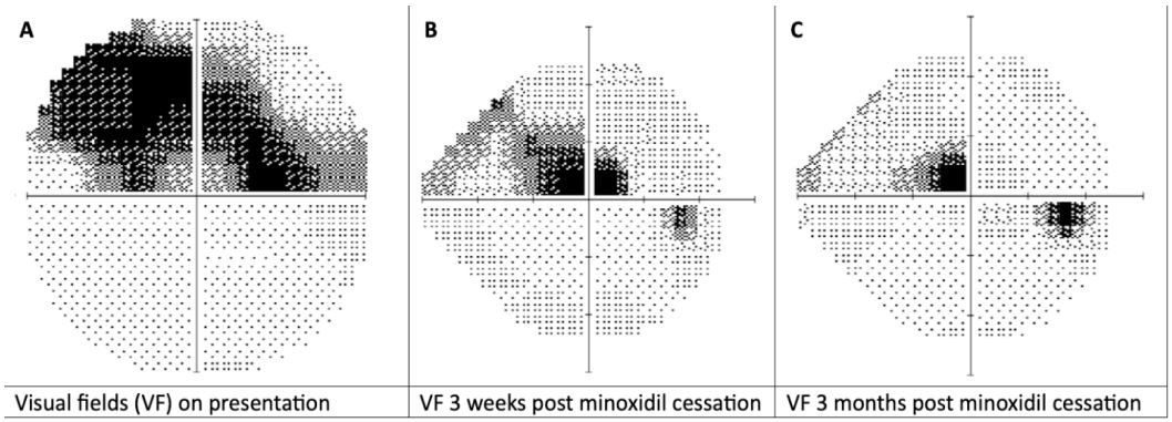
His BCVA OD improved to 20/20-1 within the first week following the cessation of minoxidil. His Visual Fields (VF) on presentation improved from near-total vision loss in the superior
visual fields (Figure 4A) to residual VF defects superior-centrally
at week 3 (Figure 4B), and to mild central VF defects at 3 months
(Figure 4C). Although both the BCVA and VFs showed improvement within the first week, fundus photography at week one
revealed worsening macular edema (Figure 5A). However, there
was significant improvement by week 3 (Figure 5B). Evaluations
after 3 months post-cessation of minoxidil showed his BCVA to
be 20/20 OU, with no other complaints.
Discussion
We present a unique case of Central Retinal Vein Occlusion
(CRVO) with secondary Cilioretinal Artery Occlusion (CLRAO) in
a patient using oral Minoxidil for androgenic alopecia. While
Minoxidil oral tablets are typically reserved for the treatment of
refractory hypertension as it functions as direct vasodilator, the
topical formulation is a popular treatment for androgenic alopecia, approved by both FDA and Health Canada [7]. Although
minoxidil is generally considered to be a safe medication, few
articles have shown systemic adverse events and ocular pathologies associated with the use of topical formulation of minoxidil
[3-6]. However, reports of CRVO with secondary CLRAO, in the
context of either topical or oral Minoxidil usage, have not been
previously documented in the literature.
The cilioretinal artery arises from the posterior ciliary artery and is susceptible to sudden occlusion due to blockage of
blood flow. CLRAO is a rare condition but most associated with
systemic vascular diseases like atherosclerosis, giant cell arteritis, and often secondary to CRVO [8-10]. Our patient was diagnosed with CRVO and secondary CLRAO, supported by fundal
examinations, Optical Coherence Tomography (OCT) findings,
and Fluorescein Angiography (FA), all indicative of territorial involvement of the central retinal vein and the cilioretinal artery
(Figures 1 & 2). Recent literature has suggested that a combined
CRVO and CLRAO could be triggered by a temporary hemodynamic blockade in the cilioretinal artery, which occurs when a
sudden increase in intraluminal pressure within the retinal capillary bed surpasses that within the cilioretinal artery [11]. This
may also explain the FA findings which showed normal arterial
filling with the cilioretinal artery.
Although arterial and venous occlusions are usually associated with several other etiological factors as mentioned above,
our patient had no other known risk factors that could predispose to such an ocular event. Nortriptyline, another medication
the patient was taking, does not have any known association with ocular pathologies according to literature review and product information, and the patient continued its usage throughout
without ocular issues. Additionally, the Naranjo adverse drug
reaction probability score was calculated to be 5, suggesting
a probable idiosyncratic drug reaction to minoxidil usage [12].
Based on clinical history and ocular findings, the use of minoxidil, the subsequent improvement in the BCVA and VFs (Figures 4
& 5) following the cessation of minoxidil, in an otherwise young
and healthy patient, this rare occurrence of combined CRVO
and CLRAO is likely due to use of minoxidil.
Conclusion
It is worth noting that while there have been reports linking
topical Minoxidil to ocular pathologies as mentioned above, to
our knowledge, there have been no previous reports regarding
ophthalmic side effect of oral minoxidil use, specifically CRVO
with secondary CLRAO. Given the rarity of such reports, further
investigation is warranted to explore the relationship between
Minoxidil use, whether topical or oral, and adverse ophthalmic
outcomes. This case underscores the necessity for increased
awareness among healthcare providers regarding the potential
ophthalmic side effects of Minoxidil, to ensure prompt recognition and management of such complications.
Declarations
Contributions: Motaz Bamakrid and Sidratul Rahman participated in preparing the manuscript equally.
Dr. Christian El-Hadad is the principal investigator, reviewed
the manuscript, provided feedback, guidance, and expertise on
the topic.
Consent: None required for case reports.
Competing interest: None.
References
- Messenger AG, Rundegren J. Minoxidil: Mechanisms of action on hair growth. Br J Dermatol. 2004; 150(2): 186-94.
- Nestor MS, Ablon G, Gade A, Han H, Fischer DL. Treatment options for androgenetic alopecia: Efficacy, side effects, compliance, financial considerations, and ethics. Journal of cosmetic dermatology. 2021; 20(12): 3759-3781. https: //doi.org/10.1111/jocd.14537
- Venkatesh R, Pereira A, Reddy NG. et al. Retinal artery occlusion as a probable idiosyncratic reaction to topical minoxidil: A case report. J Med Case Reports. 2021; 15: 493. https: //doi.org/10.1186/s13256-021-03114-8
- 4. Aktas H, Alan S, Türkoglu EB, Sevik Ö. Could Topical Minoxidil Cause Non-Arteritic Anterior Ischemic Optic Neuropathy?. Journal of clinical and diagnostic research: JCDR. 2016; 10(8): WD01-WD2. https: //doi.org/10.7860/JCDR/2016/19679.8250
- Fabio Scarinci, Paolo Mezzana, Paola Pasquini, Michelle Colletti, Andrea Cacciamani. Central horioretinopathy associated with topical use of minoxidil 2% for treatment of baldness, Cutaneous and Ocular Toxicology. 2012; 31: 157-159. DOI: 10.3109/15569527.2011.613427
- Reza Rastmanesh. Alopecia and ocular alterations: a role for Minoxidil?, Journal of Receptors and Signal Transduction. 2010; 30: 189-192. DOI: 10.3109/10799891003786234
- Sica DA, Gehr TW. Direct vasodilators and their role in hypertension management: Minoxidil. J Clin Hypertens (Greenwich). 2001; 3(2): 110-4. doi: 10.1111/j.1524-6175.2001.00455.x.
- Hayreh SS. Central retinal artery occlusion. Indian J Ophthalmol. 2018; 66(12): 1684-1694. doi: 10.4103/ijo.IJO_1446_18.
- Kumaran N, Liyanage S, Angunawela R, Asaria R. Cilioretinal artery occlusion secondary to an evolving retinal vein occlusion. Can J Ophthalmol. 2008; 43(1): 127. doi: 10.3129/i07-197.
- McDonald HM, Handzic A, Margolin E. Cilioretinal artery occlusion secondary to central retinal vein occlusion. Can J Ophthalmol. 2023 Sep 17: S0008-4182(23)00271-5. doi: 10.1016/j.jcjo.2023.08.010.
- Pinna A, Zinellu A, Serra R, Boscia G, Ronchi L, et al. Combined Branch Retinal Artery and Central Retinal Vein Occlusion: A Systematic Review. Vision (Basel, Switzerland). 2023; 7(3): 51. https: //doi.org/10.3390/vision7030051
- Liver Tox. Clinical and Research Information on Drug-Induced Liver Injury. Bethesda (MD): National Institute of Diabetes and Digestive and Kidney Diseases. 2012-. Adverse Drug Reaction Probability Scale (Naranjo) in Drug Induced Liver Injury. 2019.


